Intramedullary nailing is a well-established technique for fracture repair. This method effectively stabilizes fractures while promoting healing. nail intramedullari provides a minimally invasive option, leading to reduced surgical trauma and quicker recovery times. Surgeons often favor this approach because of its versatility.
However, the application of intramedullary nails is not always straightforward. The choice of nail size and type can greatly influence the outcome. It's crucial to consider the specific fracture pattern and the patient's unique anatomy. Sometimes, even experienced surgeons face challenges with alignment and hardware placement.
Understanding the best techniques for nail intramedullari is essential. Each method has its pros and cons, requiring careful consideration. The goal remains to achieve stable fixation and optimal healing. As techniques evolve, ongoing assessment is necessary to enhance clinical practices. Continuous learning is integral in this field.
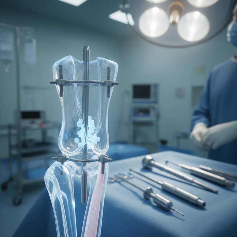
Intramedullary nails are widely used in fracture repair. They provide internal support for bone healing. These nails are inserted into the medullary cavity of long bones. This technique allows for stable fixation. Surgeons often choose intramedullary nails for their effectiveness. They minimize soft tissue disruption compared to other methods.
When using intramedullary nails, precision is crucial. Proper alignment of the nail can impact recovery. Misalignment may lead to complications. Some surgeons experience challenges in nail insertion. Selecting the correct nail size is vital. An incorrectly sized nail can cause additional issues.
Incorporating advanced imaging techniques during surgery can help. It improves accuracy in nail placement. Nonetheless, there are cases where errors occur. Surgeons must continually refine their skills. The learning curve for this technique is steep. Each procedure offers opportunities for improvement. Recognizing and addressing shortcomings can enhance future outcomes.
This chart illustrates the effectiveness of various intramedullary nail techniques in fracture repair. As shown, reamed nailing has the highest effectiveness percentage, while unlocked nailing shows the lowest effectiveness among the techniques evaluated.
Intramedullary nailing is a popular technique in orthopedic surgery. It is often used for stabilizing fractures. This technique is particularly beneficial for long bone fractures in adults. The femur, tibia, and humerus are common areas where this method is applied. The intramedullary nail helps align bones, facilitating proper healing. Surgeons choose this method because of its effectiveness in reducing post-operative complications.
Indications for using intramedullary nailing include unstable fractures or those at risk of nonunion. For example, comminuted fractures can benefit significantly from this technique. The nail provides internal support, allowing for early mobilization. Yet, it is not always the best option. Some fractures may require different surgical approaches. Misalignment can occur if the assessment is inaccurate before the procedure. Each case is unique and requires careful consideration.
Surgeons must evaluate various factors before deciding on intramedullary nailing. Patient age, bone quality, and activity level are crucial. The anatomical location of the fracture also plays a role. In some instances, using this method might lead to complications. For example, infection is a concern. Understanding the risks is essential for optimal patient outcomes.
The intramedullary nail insertion technique is a vital procedure for effective fracture repair. It plays a significant role in improving patient outcomes. Studies indicate that intramedullary nailing can achieve a union rate of up to 95% for certain fractures. This technique minimizes soft tissue damage, promoting faster recovery.
During the procedure, surgeons should begin with proper patient positioning. A clean surgical field is essential to prevent infections. The next step involves making precise incisions to access the medullary cavity. Fluoroscopy guidance ensures correct placement of the nail. Recent reports highlight that about 15% of procedures face complications such as malalignment or infection, which underscores the importance of meticulous technique.
Intraoperative assessments are crucial. Surgeons must regularly check alignment and rotation during insertion. Utilizing advanced imaging aids can help; however, each case may demand unique adjustments. Data show that even experienced surgeons encounter unexpected challenges in difficult fractures. Continuous learning and adaptation are vital for improving technique and patient safety.
Intramedullary nailing is a common technique for treating fractures, but complications can arise. According to recent studies, approximately 10-20% of patients experience complications like infection, nonunion, or malalignment post-surgery. These issues can significantly impact recovery time and overall outcomes.
Infection is a major concern. It can develop due to exposure during surgery or even from hardware. Surgical site infections occur in about 1% to 5% of cases, according to some orthopedic journals. Proper sterilization and technique are crucial. Additionally, if the bone does not heal correctly, a revision surgery may be needed, leading to increased recovery time and costs.
Malalignment is another common complication. Misplacement of the nail can result in improper healing angles, which affects limb function. Studies show that up to 15% of cases may present this issue. Early detection is key. Regular follow-ups and imaging can help identify these complications. Addressing them promptly can prevent long-term issues, but awareness of these risks is essential for both patients and surgeons.
Rehabilitation after intramedullary nail surgery is crucial for recovery. Patients often face challenges in mobility and pain management. A structured protocol helps address these issues effectively. Early weight-bearing is often encouraged, but it should be monitored closely. Patients must be aware of their limits.
Physical therapy plays a significant role in rehabilitation. Simple exercises can begin soon after surgery. These include ankle pumps and gentle knee bends. Gradual progression to more intense activities is essential. Pain can hinder progress, so patients need regular communication with their therapists. Adjustments to exercise routines may be necessary.
Setting realistic goals is important. Recovery can be unpredictable. Some patients may find it harder to regain full function than expected. This could lead to frustration. Reflecting on individual progress helps in understanding limitations. Modification of the rehabilitation plan may boost confidence and improve outcomes.
| Technique | Indications | Advantages | Rehabilitation Protocol |
|---|---|---|---|
| Closed Intramedullary Nailing | Diaphyseal fractures of long bones | Minimally invasive, less soft tissue damage | Early motion, gradual weight bearing |
| Reamed Intramedullary Nailing | Comminuted fractures, non-union cases | Improved stability, promotes healing through reaming | Progressive rehabilitation, guided by pain levels |
| Blocking Screws Technique | Spiral fractures, fracture instability | Prevention of rotation, enhanced stability | Weight bearing as tolerated, focus on range of motion |
| Static vs Dynamic Nailing | Fractures requiring different healing strategies | Flexibility to adapt to fracture type | Tailored rehabilitation protocol based on nail type |
